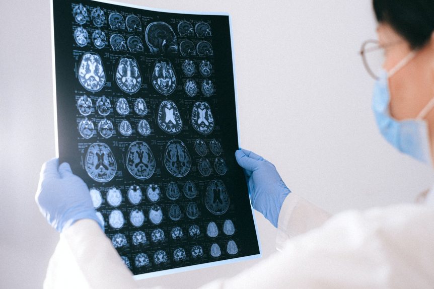
MRI, short for Magnetic Resonance Imaging, is a wonderful medical technology that provides detailed images of the internal structures of the body. Unlike X-rays or CT scans, MRI uses powerful magnets and radio waves to generate these images, making it a non-invasive and radiation-free diagnostic tool.
Understanding MRI: A Closer LookMRI is a versatile imaging technique that allows physicians to examine various parts of the body with exceptional precision. It’s a critical tool in diagnosing a wide range of conditions, from musculoskeletal issues to neurological disorders. Let’s explore how MRI sheds light on different aspects of our anatomy:
- Brain and Central Nervous System: MRI scans of the brain provide unparalleled insights into its intricate structures. This is invaluable in detecting abnormalities such as tumors, aneurysms, and multiple sclerosis lesions. Neurologists rely heavily on these images for accurate diagnoses.
- Spine and Joints: When it comes to the spine and joints, MRI is a game-changer. It helps identify issues like herniated discs, ligament tears, and arthritis. With its high-resolution images, it enables orthopedic specialists to plan surgeries and other interventions with precision.
- Abdomen and Pelvis: MRI is a go-to for assessing abdominal organs like the liver, kidneys, and pancreas. It’s especially useful in detecting tumors, cysts, and abnormalities in the gastrointestinal tract. For pelvic examinations, it provides detailed images of reproductive organs.
- Breast Tissue: In breast imaging, MRI plays a crucial role, especially for high-risk patients. It can detect early signs of breast cancer that might not be visible on a mammogram. For women with dense breast tissue, an MRI can be an essential screening tool.
- Eye Scans: MRI technology has also found applications in ophthalmology. Specifically, it is used to create detailed images of the eye and its surrounding structures. This can help an opthalmologist diagnose and monitor various eye conditions, such as optic nerve disorders and certain tumors.
- Cardiovascular System: While MRI is not the primary tool for heart imaging, it can provide essential information about heart structure and blood flow. It’s particularly useful for evaluating congenital heart defects and assessing heart function after a heart attack.
Before the Procedure:
Before your MRI, there are a few important things to keep in mind. Dressing appropriately is crucial, as metal objects can interfere with the imaging process. Just like how the nurses and medical professionals dress in comfortable scrubs to maintain hygiene and free movement, you too would likely be given a hospital gown to wear during the procedure. Make sure to inform your doctor about any metal implants or devices in your body. Additionally, avoid wearing makeup, as some formulations may contain metallic elements.
During the Procedure:During the MRI, you’ll be positioned on a comfortable table that slides into the machine. It’s essential to remain still, as any movement can blur the images. You may hear loud tapping or knocking sounds, which are normal parts of the imaging process.
After the Procedure:Once the MRI is complete, you’ll be able to resume your normal activities. However, if you were given a contrast agent, you may need to wait for a short while to ensure there are no adverse reactions. The images generated will be sent to a radiologist for interpretation.
Conclusion:MRI is a remarkable tool that has revolutionized the field of medical imaging. Its ability to provide detailed, high-resolution images of various body structures has been instrumental in diagnosing and treating a wide range of conditions. By understanding what to expect before, during, and after an MRI, patients can approach the procedure with confidence, knowing that they are taking an important step towards better health.
Although an MRI produces images almost immediately, it can take anywhere from a few days to two weeks for radiologists to review them and doctors to report back. This is why, as the infographic states, it is a good idea to ask how long that report will take before you leave. For all the MRI details, please continue reading.
- Best learning toys for children as they age - July 19, 2023
- Luxury yacht charter vs. standard yacht charter: Which is right for you? - February 7, 2023
- Comfortable Shoes for Being on Your Feet All Day - January 10, 2023

Like It? Share It!